What is a magnetic resonance imaging (MRI) scan?

1
MRI (magnetic resonance imaging) is a medical imaging technique used to visualise internal body structures, such as the brain, spinal cord, and joints, as well as internal organs such as the heart, lungs, liver, and kidneys. This method uses magnetic fields and radio waves to create detailed images of tissue.
2
During the MRI scan, the patient is positioned on a special table, which then moves between a large magnet and sources of radio waves. The MRI machine is similar to a tunnel surrounded by a magnetic field. The magnetic field causes hydrogen atoms in the body to react, emitting signals that are then used to create an image. MRI scans are non-invasive and do not use ionising radiation, so they are a safe method of tissue examination.
3
In general, MRI scans are an important tool in the diagnosis and monitoring of many medical conditions due to their ability to provide detailed and versatile images of internal parts of the body.

Please watch the video!

In general, MRI scans are an important tool in the diagnosis and monitoring of many medical conditions due to their ability to provide detailed and versatile images of internal parts of the body.

How do I prepare for an MRI scan?

1

Fill out the questionnaire and bring it with you to the scan:

2

Please be sure to arrive at least 15 minutes prior to the scan!

3

When going to the scan, wear comfortable, active clothing. Please note that metal objects containing magnetic metals (e.g. iron) must not be brought into the scanning room.
Jewellery must not be taken into the scanning room. Jewellery, wallets, credit cards, and other items that might interfere with the scanning process can be placed in a security cabinet. Before the scan, you may be asked to drink a contrast agent or, if needed, it will be injected intravenously. Be sure to inform the radiology technician about any other medical conditions you may have, as intravenous injections may be contraindicated for certain diseases.Inform the radiology technician of any allergies or allergic reactions to medications. Always tell the radiology technician if you might be pregnant. Although there have been no adverse effects on the foetus found with MRIs, it should still be avoided in the first trimester of pregnancy. At the same time, it is possible that the benefits of the scan outweigh the unlikely risks. If you suffer from claustrophobia, ie a fear of enclosed spaces (such as lifts), please be sure to inform the staff. It is not permitted to enter the scanning room with a pacemaker. It is not possible to perform an MRI scan on patients with a pacemaker. However, there are now pacemakers that can withstand the magnetic field, but an electrophysiologist must confirm that the scan can be performed and, if necessary, the pacemaker must be reconfigured to a mode that is suitable for an MRI.

If you have undergone previous surgeries and have any implanted foreign bodies such as joint prostheses or heart valves, please be sure to inform our staff. Some of these may contain materials that should not be brought into the scanning room. However, most modern prostheses are made of materials compatible with MRI examinations.
IMPORTANT!

We cannot perform an MRI scan in the following cases:

1
Less than six weeks has passed since the implantation of the pacemaker, cardioverter-defibrillator or DBS electrodes.
2
You have an abandoned or epicardial electrode.
3
The pacemaker battery is low.
4
You have a prosthetic heart valve or aneurysm clip that contains metal and was implanted before 1996.
5
You have a hearing implant (non-removable), except for a hearing implant whose suitability for an MRI is certified by the manufacturer (MR safe, MR conditional) and which has a document describing the conditions under which an MRI is allowed.
6
You have a metal fragment in your eye, eye socket, head, or spinal region.
After the MRI scan, you will be able to return to your normal activities. The doctor will evaluate the images and inform you of the results in writing on your Health Portal page.

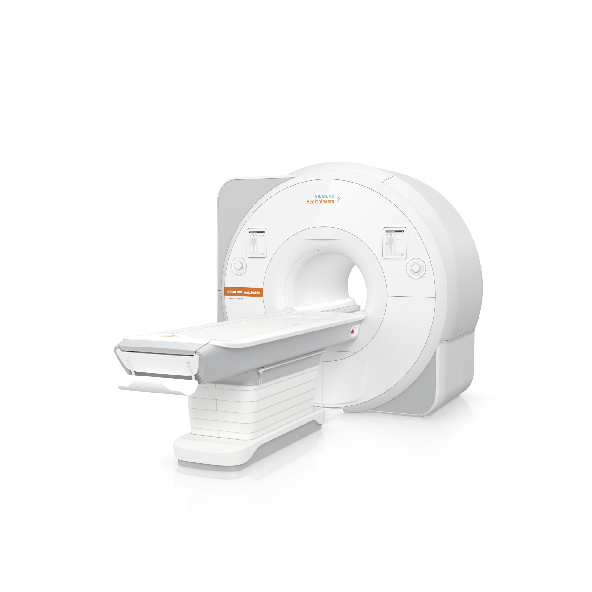
How is the MRI scan performed?

A magnetic resonance imaging (MRI) scan is a series of steps that produce detailed images of the inside of the body. Here is a general overview of what happens during an MRI scan:
Preparation
Before the scan, you may be asked to remove jewellery, metal objects, and other items that may interfere with the magnetic field. You may also be asked about your health condition to make sure the scan is safe for you. If you are going to be injected with contrast, you may be asked about allergies and previous reactions to contrast agents.
Clothing
You will be asked to undress and change into comfortable clothing provided by the clinic. This ensures that your clothing does not contain metals that could affect the quality of the MRI scan.
Positioning
You will be positioned on the MRI examination table, which will then move inwards into the tube of the MRI machine. It is very important that you remain motionless while the information is being collected. The information is collected in stages. One collection round can last from a couple of seconds to ten minutes. While the information is being collected, you will hear a rumbling or tapping sound. During the break between the stages of information collection, you can relax. When examining certain parts of the body, it is possible to position the patient with their head outside the tunnel. Unfortunately, this is not possible with all scans. Sometimes supports, cushions or Velcro straps are placed around the area of examination. These are used to keep the part of the body in the most comfortable position. Plastic frames or wraps may be placed over the area being examined to enhance signal reception.
Scanning
The MRI scanner generates a strong magnetic field and directs radio waves towards the body’s internal structures. The magnetic field causes hydrogen atoms in the body to react, emitting signals that are then used to create the image. The scan can take anywhere from a few minutes to an hour, depending on the area being examined and the type of images needed.
Contrast agent injection (if necessary)
In some cases, a contrast agent may be intravenously injected during the scan to help visualise certain parts of the body or tissue. This may be necessary, for example, when examining blood vessels or tumours. If a contrast agent is to be used, the radiology technician will insert an IV (intravenous) cannula. The IV cannula is placed on the forearm and a tube is attached to it, in order to allow the contrast agent to be injected without moving the examination table. The scan is controlled from a computer that is outside the scanning room.
Finishing the screening
After the scan, you will assisted out of the MRI scanner. You can put your clothes back on. If you were injected with a contrast agent, you may be advised to monitor yourself for any potential reactions for a period of time.

What will I feel during the scan?

It is important to note that MRI scans are generally safe and are well tolerated by most people. If you experience discomfort or anxiety during the examination, please make sure to inform our radiology technician. We strive to make your visit as comfortable as possible by providing pillows, towels, headphones, etc. Your well-being is our priority. During the MRI (magnetic resonance imaging) scan, patients may experience various sensations, most of which are typically mild and tolerable. Here are some of the feelings and other things you may experience during an MRI scan:
Noise
The MRI scanner produces a loud noise that can sound like knocking or rattling. We provide headphones and/or earplugs to reduce the impact of noise.
Narrowness
The MRI machine has a wide opening, but is still in the shape of a tube, which can make you feel that the space is small or cramped. This can cause a slight feeling of anxiety in some people. If you find it difficult to continue the screening, there is an emergency button you can press.
Immobility
Remaining motionless is important for getting a clear image. Some people find it difficult to stay still for long periods of time.
Warmth
Some patients may feel a slight sensation of warmth during an MRI scan, especially when a contrast agent is injected.
Sensitivity to contrast agent injection
If a contrast agent is used during the MRI scan, the injection may cause a brief sensation of heat or a slight tingling.
IV cannula
The insertion of the cannula may be a little uncomfortable at first, and there may be some swelling when the cannula is removed. During the injection of the contrast agent, there is often a cold sensation in the arm.
Exposure to a magnetic field
After the scan
After an MRI scan, you can return to your normal day. A recovery period will only be necessary if sedatives have been used. Very rarely, nausea or an allergic rash may occur after contrast agent injection. Breastfeeding is not recommended for two days after the administration of the contrast agent.

Book your MRI scan now

Cranfeld Clinic OÜ
Veerenni tn 53a Tallinn Harjumaa 10138
Registry code: 16385217

    NAME *

    EMAIL *

    PHONE *

    TEXT *

    The booking system is currently under development.
    Please contact us at mrt@cranfeld.ee or +372 5303 6257.